| What it treats | AVMs, venous and lymphatic malformations, selected hemangiomas |
|---|---|
| Anaesthesia | Local with sedation, or general anaesthesia (especially for children) |
| Entry point | Needle puncture in the groin or wrist, or direct needle into the lesion |
| Time taken | Usually 1–3 hours |
| Hospital stay | Usually same day to 1–2 days |
| Sessions | Often more than one for large lesions |
This page is about AVMs outside the brain. If you have been told you have an AVM in the brain or spinal cord, ask your neurologist about assessment by a neuro-interventional and neurosurgical team.
Who may need AVM embolisation
AVM embolisation is for AVMs that cause problems, such as bleeding, ulcers, pain, fast growth or strain on the heart. A quiet AVM that causes no trouble is often only watched.
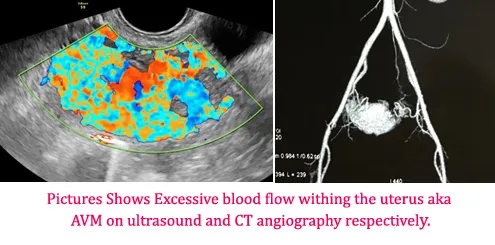
An AVM (arteriovenous malformation) is a tangle of abnormal vessels where arteries connect straight to veins. Outside the brain, it usually shows as a warm, pulsing lump in an arm, leg, the face or the pelvis. Read what an AVM outside the brain is, its stages and how it is diagnosed.
Embolisation may be advised if your AVM:
- Bleeds, from the skin or, in a uterine AVM, as heavy vaginal bleeding
- Has caused a sore (ulcer) or skin breakdown
- Hurts, or limits use of the hand, foot or limb
- Keeps growing
- Is large enough to strain the heart
- Changes the shape of the face in a way that troubles you
How it works: the nidus and why it must be closed
Embolisation works by closing the nidus, the knot at the centre of the AVM where arteries join veins. Closing the nidus is what stops the AVM, not blocking the arteries that feed it.
Think of the nidus as a busy junction where many roads meet. If you block only the roads leading in, traffic finds new routes. In the same way, if only the feeding artery is tied off or blocked, the AVM recruits new small arteries and often grows back, sometimes worse. This is why surgeons no longer tie off feeding arteries on their own.
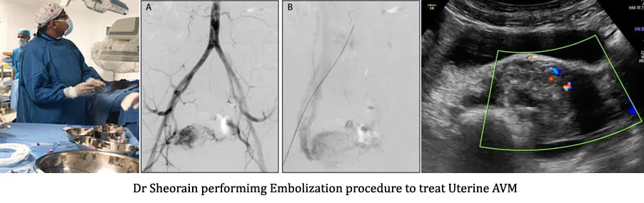
In embolisation, the agent is placed right into the nidus. It fills and seals the abnormal channels. The healthy vessels around it are left open.
Agents used
Several agents are used to close an AVM, and the choice depends on the AVM’s size, site and flow. Many treatments use more than one.
| Agent | How it works | When it may be used |
|---|---|---|
| Ethanol (pure alcohol) | Damages the lining of the abnormal vessels so they scar and close | Some AVMs where lasting closure of the nidus is the goal; needs care near skin and nerves |
| Liquid embolic agent | A liquid that sets into a soft solid inside the vessels, filling the nidus | Many AVMs reached through a catheter |
| Glue | Sets almost at once on contact with blood, sealing the channel | Selected AVMs; fast-flow channels |
| Coils | Tiny metal coils that pack a vessel and cause a clot | Large draining veins or fast channels, often with another agent |
| Plugs | Small expanding devices that block a larger vessel | Large single channels |
What happens, step by step
AVM embolisation is done in a cath lab or interventional suite, guided by X-ray and sometimes ultrasound. Here is what usually happens:
- Planning scans. An MRI or CT angiogram maps the AVM. Doppler ultrasound shows the flow.
- Anaesthesia. Depends on the site and agent: local anaesthesia with sedation, or general anaesthesia. Ethanol treatment is usually done under general anaesthesia.
- Access. A thin tube (catheter) is passed into the artery, often through a needle puncture in the groin or wrist. Some AVMs are reached instead with a fine needle placed straight into the nidus through the skin.
- Mapping. Dye pictures (angiography) show the nidus exactly.
- Embolisation. The agent is injected slowly into the nidus, with pictures taken as it goes.
- Checks. Further dye pictures show how much of the AVM is closed.
- Finish. The catheter is removed and the puncture is pressed or closed.
The procedure usually takes one to a few hours, depending on the size and site. Many people go home the same day or the next day.
Why treatment is often staged
Large AVMs are usually treated in stages, over two or more sessions some weeks or months apart. This is planned, not a sign that the first session failed.
Staging protects the skin, nerves and healthy tissue around the AVM, because closing too much at once can cause heavy swelling or damage the skin. After each session, the area settles, and a scan shows what is left. The next session targets the remaining nidus.
Small AVMs may need only one session. Your specialist plans the number after the first scans.
Recovery
Swelling and soreness are normal for some days after AVM embolisation, and they are expected as the AVM closes. Most people recover at home.
- Swelling. Usually worst in the first 2 to 3 days, then settles over 1 to 2 weeks. Keeping the limb raised helps.
- Pain. Treated with tablets. Tell the team if pain gets worse instead of better.
- Skin care. Watch the skin over the AVM. Report darkening, blistering or a new sore.
- Activity. Light activity in a few days; avoid heavy use of the treated limb for the period you are advised.
- Follow-up. A check-up and scans after some weeks to see how much of the AVM has closed.
Risks
AVM embolisation is an established treatment, but it carries risks, and these depend a lot on where the AVM is. Possible problems include:
- Swelling and pain, which are common and usually settle
- Skin damage, such as blistering, a sore or, rarely, loss of skin over the AVM
- Nerve injury, causing numbness or weakness, usually temporary
- Non-target embolisation, where the agent reaches a normal vessel, which can harm nearby tissue
- Puncture-site problems such as bruising
- Recurrence, where the AVM regrows over time
Who it is not right for
AVM embolisation is not right for every AVM. It may not be the right choice if:
- The AVM is quiet (Stage I) and causes no symptoms. Watching is often safer.
- The nidus sits right next to a vital nerve or structure, where the risk outweighs the benefit
- You are pregnant. Treatment is usually delayed until after delivery unless there is bleeding
- Surgery alone is the better option for a small, easily reached AVM. A plastic or vascular surgeon may advise this
Dr. Sheorain will tell you plainly if he thinks watching or another treatment suits you better.
Your first visit
At your first visit, Dr. Sheorain examines the AVM, reviews your scans and explains whether embolisation suits you, how many sessions it may take and what the risks are for your AVM’s site.
Bring any Doppler, MRI or CT reports with the images, and photos showing how the lump has changed. Photos of patients are used on this site only with written consent.
Medanta Vascular OPD: Floor 5, Vascular OPD, Medicity, Islampur Colony, Sector 38, Gurugram, Haryana 122001. Monday to Saturday, 9:00 AM to 5:30 PM. See how to reach the Vascular OPD and Dr. Virender Sheorain’s training. Learn what embolisation is and what interventional radiology is. Related conditions: vascular malformations and hemangioma.
Common questions about AVM and hemangioma embolization
Is AVM embolisation a surgery?
No open surgery is involved. The vessels are blocked from inside through a catheter or a needle, guided by imaging.
How many sessions will I need?
Small AVMs may need one session; larger ones often need several. Your specialist plans this after the scans.
What is the success rate of AVM embolisation?
It depends on the AVM's size, site and type. Your specialist can explain what is realistic for your AVM.
Is AVM embolisation painful?
You will usually have anaesthesia or sedation during it. Swelling and soreness afterwards are common and are treated with medicines.
Can an AVM come back after embolisation?
Yes, it can regrow, which is why follow-up scans are needed.
Sources
This page follows these patient resources and guidelines:
- Vascular malformations (RadiologyInfo, RSNA & ACR)
- Arteriovenous malformations (MedlinePlus, US National Library of Medicine)
Related
AVM (Arteriovenous Malformation)
Abnormal tangles between arteries and veins.
Learn more →Hemangioma & Birthmarks
Blood-vessel birthmarks in babies, children and adults.
Learn more →Vascular Malformations & Birthmarks
Abnormal blood vessels and blood-vessel birthmarks, in adults and children.
Learn more →
Who performs this procedure
Dr. Virender Sheorain, Director, Peripheral Vascular & Endovascular Sciences, Medanta – The Medicity, Gurugram. Trained at AIIMS New Delhi and in three fellowships in the USA.
Read Dr. Sheorain's profile →This is general information. Please consult the doctor for advice about your condition.