What is a vascular malformation?
A vascular malformation is a group of blood vessels or lymph vessels that formed in the wrong way before birth, and it can affect people of any age. It is not a cancer and it is not caused by anything the mother did in pregnancy.
Some are visible at birth as a birthmark or a soft lump. Others stay hidden for years and only show up in childhood, the teenage years or adulthood, when they start to swell or hurt. They grow slowly along with the person. They can get bigger faster during puberty, pregnancy, or after an injury.
Malformations can be found almost anywhere: the skin, the muscles of an arm or leg, the face, the pelvis or the organs.
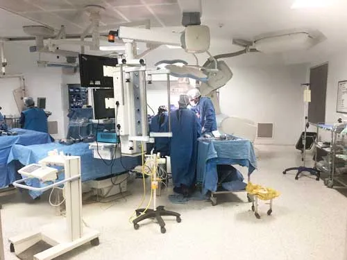
Types: slow-flow and fast-flow
Doctors group vascular malformations by how fast blood flows through them, because flow decides the treatment. Slow-flow types are more common; fast-flow types need closer care. This follows the international classification used by vascular anomaly specialists (ISSVA classification).
| Type | How it looks or feels | Flow | Main test | Usual treatment | Who usually leads |
|---|---|---|---|---|---|
| Venous malformation | Soft, bluish, squashy lump; gets bigger when the limb hangs down or with exercise; may ache | Slow | MRI, Doppler ultrasound | Sclerotherapy (injection that closes the abnormal veins); compression | Interventional radiologist |
| Lymphatic malformation | Soft swelling or cysts filled with clear fluid; may suddenly swell with infection | Slow (no blood flow) | MRI, ultrasound | Sclerotherapy; sometimes surgery | Interventional radiologist, with paediatric or plastic surgeon |
| Capillary malformation (port-wine stain) | Flat pink, red or purple birthmark | Slow | Examination | Laser | Dermatologist |
| Arteriovenous malformation (AVM) | Warm lump, may pulse or buzz; skin may be red; can bleed | Fast | Doppler, MRI, angiography | Embolisation; sometimes surgery | Interventional radiologist |
Some people have a mix of types in the same area.
Is it a hemangioma or a malformation?
A hemangioma is a growth that appears after birth, while a malformation is present from birth and grows with the person. The two words are often mixed up, even on scan reports.
- Infant hemangioma. A red, raised lump that appears in the first weeks of life, grows fast for some months, then slowly shrinks over years. Most need no treatment, or are treated with medicines by a paediatrician or dermatologist.
- Vascular malformation. Present from birth (even if not seen), does not shrink on its own, and grows slowly with the person.
Many lumps in adults that are called a “hemangioma” are actually venous malformations. The difference matters, because the treatment is different. Read about hemangioma of the skin and soft tissue, and about liver hemangioma, which is a different condition found in the liver.
Symptoms by location
Symptoms depend on where the malformation is and what type it is. Common patterns are:
- In the leg or arm: a soft swelling that gets bigger and aches after standing, walking or exercise; heaviness; visible veins; sometimes small hard lumps (tiny clots) that are tender
- In a muscle: pain with use, and swelling that comes and goes
- On the face or lips: a soft swelling or colour change, which can affect appearance and sometimes eating or speech
- In the pelvis: pelvic pain or heaviness, bleeding, or varicose veins in the area
- AVM anywhere: a warm lump that may pulse, red or dark skin, ulcers or bleeding
If the lump changes quickly, starts to bleed or becomes very painful, see a specialist soon.
Is a vascular malformation dangerous?
Most vascular malformations are not life-threatening, but they can cause pain, swelling, bleeding or problems with appearance and movement. A few need closer care:
- Fast-flow malformations (AVMs) can bleed and grow over time, and need regular follow-up.
- Large malformations near vital structures, such as the airway or eye, need careful planning.
- Large venous malformations can form small clots inside them, which cause pain. In some people they affect the body’s clotting, which is checked before treatment.
A specialist review with an MRI tells you which type you have, and how closely it needs watching.
How it is diagnosed
Diagnosis usually takes a clinical examination and one or two scans. The right tests depend on the type:
- Doppler ultrasound shows whether blood flows fast or slowly, and how deep the malformation goes. It is quick and uses no radiation.
- MRI is the main test. It shows the full size of the malformation, which tissues it involves, and its type.
- Angiography (an X-ray picture of the arteries with dye) is used for fast-flow malformations, often at the start of treatment.
- Blood tests sometimes check clotting before treatment of large venous malformations.
Treatment options
Many malformations can be treated without open surgery, and the choice depends on type, size and symptoms. Several sessions are common.
- Watch and wait. Small malformations that cause no trouble may need no treatment, only follow-up.
- Compression. Compression stockings or sleeves can ease ache and swelling in the limbs.
- Sclerotherapy (for slow-flow types). Through a fine needle, under ultrasound or X-ray guidance, a medicine is injected into the abnormal vessels. It irritates their lining so they close and shrink over weeks.
- Embolisation (for fast-flow types). Through a needle puncture, a thin tube is guided to the AVM, and materials are placed to block the abnormal connections. Read about AVM and AVM embolisation, and how embolisation works.
- Laser. For capillary malformations (port-wine stains) on the skin, done by dermatologists.
- Surgery. In selected cases, surgeons remove part or all of a malformation, sometimes after embolisation to reduce bleeding. Dr. Sheorain does not perform this surgery.
Most malformations are controlled rather than removed completely. The aim is to relieve symptoms. Some people need treatment again over the years.
When another specialist leads
Some malformations are best treated by another specialist, and Dr. Sheorain will refer you when that is the right step.
- Brain AVMs are usually managed by neuro-interventional specialists and neurosurgeons.
- Port-wine stains on the skin are treated by dermatologists with laser.
- Infant hemangiomas are usually managed by paediatricians and dermatologists.
- Malformations near the airway or eye are planned with ENT, eye or plastic surgery teams.
When to see a specialist
See a vascular specialist if you or your child has:
- a soft lump or birthmark that is growing, swelling or getting painful
- a lump that gets bigger when the limb hangs down or after exercise
- a warm lump that pulses or buzzes
- bleeding from a birthmark or lump
- repeated tender, hard spots inside a known malformation
- a “hemangioma” in an adult that has never shrunk
Go to the emergency department now for heavy bleeding that does not stop with firm pressure, or swelling that affects breathing or swallowing.
Your first visit
At your first visit, Dr. Sheorain examines the lump and reviews any scans, then explains which type it is likely to be.
- Bring: any ultrasound or MRI reports and images, photos showing how the lump has changed over time, and previous treatment records.
- Check: examination and a Doppler scan if needed; an MRI if there is none.
- Plan: watching, compression, sclerotherapy, embolisation or referral, with the reasons explained.
Medanta Vascular OPD: Floor 5, Vascular OPD, Medicity, Islampur Colony, Sector 38, Gurugram, Haryana 122001. Monday to Saturday, 9:00 AM to 5:30 PM. See how to reach the Vascular OPD and Dr. Virender Sheorain’s training.
Common questions about vascular malformations
Can a vascular malformation be treated without surgery?
Many can. Slow-flow malformations are often treated with sclerotherapy injections and fast-flow ones with embolisation, both through needle punctures. Several sessions may be needed.
Is a vascular malformation dangerous?
Most are not life-threatening but can cause pain, swelling or bleeding. Fast-flow malformations need closer follow-up. A specialist review with an MRI tells you which type you have.
Do vascular malformations grow?
They are present from birth and grow with the person. They can enlarge during puberty, pregnancy or after an injury.
Which doctor treats vascular malformations?
An interventional radiologist usually leads treatment, often with dermatology, plastic surgery or paediatrics depending on the location. Brain malformations are usually managed by neuro specialists.
Is a venous malformation the same as a hemangioma?
No. A hemangioma is a growth that usually appears after birth, and in babies it often shrinks on its own. A venous malformation is present from birth and does not shrink. Adults are sometimes told they have a hemangioma when it is actually a venous malformation.
Sources
This page follows these patient resources and guidelines:
- Vascular malformations (RadiologyInfo, RSNA & ACR)
- ISSVA classification of vascular anomalies (International Society for the Study of Vascular Anomalies)
Explore this section
AVM (Arteriovenous Malformation)
Abnormal tangles between arteries and veins.
Learn more →Hemangioma & Birthmarks
Blood-vessel birthmarks in babies, children and adults.
Learn more →AVM & Hemangioma Embolization
Closes abnormal blood vessels from inside, for adults and children.
Learn more →
About the reviewing doctor
Dr. Virender Sheorain is Director, Peripheral Vascular & Endovascular Sciences, Medanta – The Medicity, Gurugram. He trained at AIIMS New Delhi and completed three fellowships in the USA.
Read Dr. Sheorain's profile →This is general information. Please consult the doctor for advice about your condition.