| What it treats | Prevents pulmonary embolism when blood thinners can't be used |
|---|---|
| Anaesthesia | Local anaesthesia; sedation if needed |
| Entry point | Needle puncture in a neck or groin vein |
| Time taken | Placement: about 30–60 minutes; removal: 30 minutes to a few hours |
| Hospital stay | Usually same day or 1 night |
| Back to routine | Usually 1–2 days |
Emergency: Sudden breathlessness, chest pain, coughing up blood or sudden swelling of one leg can mean a blood clot. Go to the nearest emergency department now.
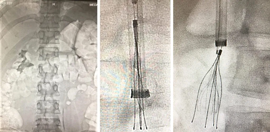
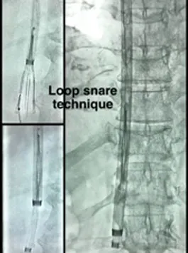
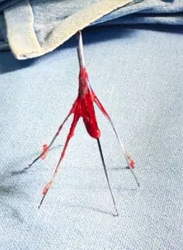
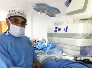
What is an IVC filter, and why was yours placed?
An IVC filter is a small metal device placed inside the body’s main lower vein to catch blood clots before they reach the lungs. The inferior vena cava (IVC) is the large vein in the belly that carries blood from the legs and lower body back to the heart.
The filter looks a little like a tiny umbrella frame or cage, made of thin metal legs that meet at a point. The legs have small hooks that hold it against the vein wall. If a clot breaks off from a leg vein, the filter traps it, so it does not travel to the lungs and cause a pulmonary embolism (a clot in the lungs).
Filters are usually placed for people who have a DVT (deep vein thrombosis) or a pulmonary embolism and:
- cannot take blood thinners, for example because of bleeding, recent surgery or an injury
- had a new clot even while on blood thinners
- needed short-term protection around a major operation or injury
There are two kinds. Retrievable filters are designed to be taken out when they are no longer needed. Permanent filters are meant to stay. Your discharge summary or the hospital that placed it can usually tell you which kind you have.
Should your filter come out? Why timing matters
A retrievable filter is meant to be temporary, and once your clot risk has passed or you can safely take blood thinners, it is worth asking a specialist whether yours should come out. The US Food and Drug Administration (FDA) advised in 2014 that doctors consider removing retrievable filters as soon as protection from a lung clot is no longer needed (FDA safety communication).
The reason is that a filter left in for a long time can cause problems in some people, such as:
- the filter tilting in the vein
- its legs pushing into or through the vein wall
- a piece of the filter breaking off
- a clot forming in or around the filter
The length of time a filter has been in the body is called its dwell time. A longer dwell time can make removal more complex, because the vein wall may have grown over the hooks. More complex does not mean impossible. It means the specialist needs to plan carefully.
Placed months or years ago, or told it can’t be removed?
Many filters that have been in place for months or years can still be removed, but each one has to be checked first. If you were told earlier that your filter cannot come out, a fresh review with up-to-date imaging is reasonable. Some filters are still safer left in place, and you will be told that honestly.
Before deciding, a CT scan or ultrasound is checked for:
- The filter type: retrievable or permanent, and which brand if known
- Tilt: whether the filter sits straight or leans to one side
- Embedded hooks: whether the hook or legs have grown into the vein wall
- Clot in the filter: a large trapped clot changes the plan
- The veins around it: whether the IVC and leg veins are open
When a standard removal is not enough, there are advanced methods. In plain words, these use different tools passed through the same neck puncture to free the filter gently from the vein wall before it is pulled into a tube.
A filter placed at another hospital can be reviewed in the same way. Bring whatever records you have.
| Your situation | What usually happens | Next step |
|---|---|---|
| Clot risk has passed and the filter is retrievable | Removal is usually advised | Book an OPD visit with your reports |
| You still cannot take blood thinners and your clot risk is high | The filter usually stays for now | Review again when your situation changes |
| Filter tilted, or hooks grown into the vein wall | Removal may still be possible with advanced methods | CT scan and specialist review |
| Permanent filter type | Usually left in place | Ask whether any follow-up is needed |
| You do not know what filter you have | Not known until records or imaging are checked | Bring your discharge summary and any scans |
Who should NOT have the filter removed (yet)?
Removal is not right for everyone, and for some people the safer choice is to leave the filter where it is. You may need to wait, or keep the filter, if:
- you still have a high risk of a lung clot and cannot take blood thinners
- there is a large clot trapped inside the filter
- you have a permanent filter that was not designed to come out
- when the risks of removal are higher than the benefit, which is explained after imaging (Kuyumcu and Walker, 2016)
Never stop or change blood thinners to get ready for removal unless your doctor tells you to.
How removal is done, step by step
Filter removal is a minimally invasive procedure done through a needle puncture, usually in the neck, without an open cut. Here is what usually happens:
- Numbing. You lie on your back. The skin over a vein in the neck, or sometimes the groin, is numbed with local anaesthesia. Light sedation is given if needed.
- Needle puncture. A thin tube is passed into the vein. There is no cut.
- Picture of the vein. Dye and X-ray (a cavogram) show the filter and check for clots.
- Catching the filter. A wire loop called a snare grabs the hook at the top of the filter.
- Closing it into a tube. A wider tube (sheath) slides over the filter, folding its legs so it comes away from the vein wall.
- Removing it. The filter is pulled out through the neck puncture, and pressure is applied.
A simple removal usually takes under an hour. Complex removals take longer. Many patients go home the same day.
How filter placement is done
Placing a filter is a short procedure through a needle puncture in the neck or groin, under local anaesthesia. A thin tube is guided to the IVC with X-ray, and the folded filter is pushed out so it opens and grips the vein wall. Most people feel pressure rather than pain. Dr. Sheorain also places filters when they are needed.
Recovery and blood thinners afterwards
Most people return to light daily activity within a day or two after removal, with a little soreness at the puncture site. Avoid heavy lifting for a short time, as advised.
Whether you need blood thinners after removal depends on why the filter was placed and your clot risk today. Many people do need them for a period. Your treating doctor and the specialist decide this together. Do not change any medicine on your own.
IVC filter or blood thinners?
Blood thinners are the main treatment for DVT and pulmonary embolism, and a filter is used only in specific situations when blood thinners cannot be used or have not worked.
| Blood thinners | IVC filter | |
|---|---|---|
| When used | First treatment for most DVT and lung clots | When blood thinners cannot be used, or a clot formed despite them |
| How it works | Stops the clot growing and lowers the chance of new clots | Catches clots travelling from the legs before they reach the lungs |
| Limits | Can cause bleeding; needs regular use | Does not treat the clot itself or stop new clots forming; retrievable filters are meant to come out later |
Source: Kuyumcu and Walker, 2016.
Risks
IVC filter removal is generally a low-risk procedure, but no procedure is free of risk. Possible problems include:
- Bruising or bleeding at the puncture site
- The filter cannot be removed at that attempt, so it stays in place or another attempt is planned
- Rarely, injury to the vein during removal
- Rarely, a piece of the filter stays behind
In published series, a standard removal succeeds in about 8 to 9 out of 10 cases, and advanced methods can remove many of the rest (Kuyumcu and Walker, 2016).
When to get emergency care
Emergency: Go to the nearest emergency department now if you have sudden breathlessness, chest pain, coughing up blood, a fast heartbeat with dizziness, or sudden painful swelling of one leg. These can be signs of a clot.
Your first OPD visit
At your first visit, Dr. Sheorain checks what filter you have, why it was placed and whether it can safely come out.
- Bring: the discharge summary that mentions the filter, the date it was placed, the hospital, any CT scans, and your current medicines.
- Talk: why the filter was placed and whether that reason still applies.
- Imaging: a CT scan or ultrasound if there is no recent one.
- Plan: removal now, removal later, or leaving it in, with the reasons explained.
Medanta Vascular OPD: Floor 5, Vascular OPD, Medicity, Islampur Colony, Sector 38, Gurugram, Haryana 122001. Monday to Saturday, 9:00 AM to 5:30 PM. See how to reach the Vascular OPD, Dr. Virender Sheorain’s training and credentials, DVT clot removal and other vein problems.
Videos: IVC filter placement and removal

Common questions about IVC filter placement and removal
Is IVC filter removal a major surgery?
No. It is a minimally invasive procedure done through a needle puncture, usually in the neck, under local anaesthesia. There is no open cut, and many patients go home the same day. Complex or long-standing filters can take longer.
Can an IVC filter be removed after many years?
Often yes, but it depends on the filter type and how it sits in the vein. A CT scan or ultrasound helps the specialist decide whether removal is safe and which technique to use. Some filters are safer left in place.
Is IVC filter placement or removal painful?
It is usually done under local anaesthesia, sometimes with light sedation, so most patients feel pressure rather than pain. A little soreness at the puncture site for a day or two is common.
Do I still need blood thinners after an IVC filter?
That depends on why the filter was placed and your current clot risk. Many patients do need blood thinners for a period; your treating doctor and the specialist decide together. Do not stop any medicine on your own.
Why should a filter that is not needed be taken out?
Retrievable filters are designed as temporary devices. Health regulators advise removing them once they are no longer needed, because leaving them long term can lead to problems with the filter or vein (FDA). A specialist can check whether yours should come out.
Sources
This page follows these patient resources and guidelines:
- Inferior vena cava filter retrievals, standard and novel techniques (Kuyumcu and Walker, 2016) (Cardiovascular Diagnosis and Therapy (NIH PMC))
- IVC filters: placement and removal (RadiologyInfo, RSNA & ACR)
- Venous thromboembolism (NHLBI, US National Institutes of Health)
Related
Deep Vein Thrombosis (DVT)
A blood clot in a deep leg vein, often with sudden swelling and pain.
Learn more →Pulmonary Embolism
A clot that has travelled to the lungs.
Learn more →DVT Clot Removal
Dissolves or removes a large leg clot through a thin tube.
Learn more →
Who performs this procedure
Dr. Virender Sheorain, Director, Peripheral Vascular & Endovascular Sciences, Medanta – The Medicity, Gurugram. Trained at AIIMS New Delhi and in three fellowships in the USA.
Read Dr. Sheorain's profile →This is general information. Please consult the doctor for advice about your condition.